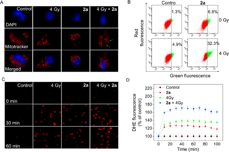
Figure 3.
Synergistic effect of 2a and X-rays on induction of mitochondrial dysfunction and ROS generation. (A) Fluorescent micrographs of mitochondrial fission induced by 2a (1 μM) in the absence or presence of X-rays (4 Gy). The photomicrographs were detected using Mitotracker and DAPI costaining. (B) Flow cytometry analysis of the changes in ΔΨm on HeLa cells treated with 2a (1 μM) without or with X-ray treatment. (C) Fluorescence images of intracellular ROS detected by DHE probe on HeLa cells. (D) Fluorescence microplate readings for intracellular ROS measured by DHE probe on HeLa cells.

