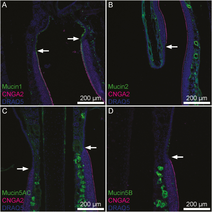
Figure 1.
Transitions between OE and RE in mouse nasal tissue. CNGA2 (magenta), a marker specific to neurocilia of OSNs indicates portions of OE. Visible portions of OE and RE are broadly representative of these tissues. (A) Mucin 1 (green) is present uniformly across a continuous layer of sustentacular cells within the OE whereas in the RE scattered individual surface epithelial cells express it. (B) Mucin 2 (green) is expressed uniformly within lamina propria connective tissue below both OE and RE. (C) Mucin 5AC is expressed by submucosal glands and to a lesser degree within lamina propria connective tissue below both OE and RE. The density of positively staining glands was much higher in the OE. (D) Mucin 5B was expressed intensely by submucosal Bowman’s glands within the OE, but within the RE goblet cells expressed mucin 5B. Arrows denote the transition from RE to OE. Also, this transition is noticeable as the thickness of the epithelial layer changes from thick to thin from OE to RE.

