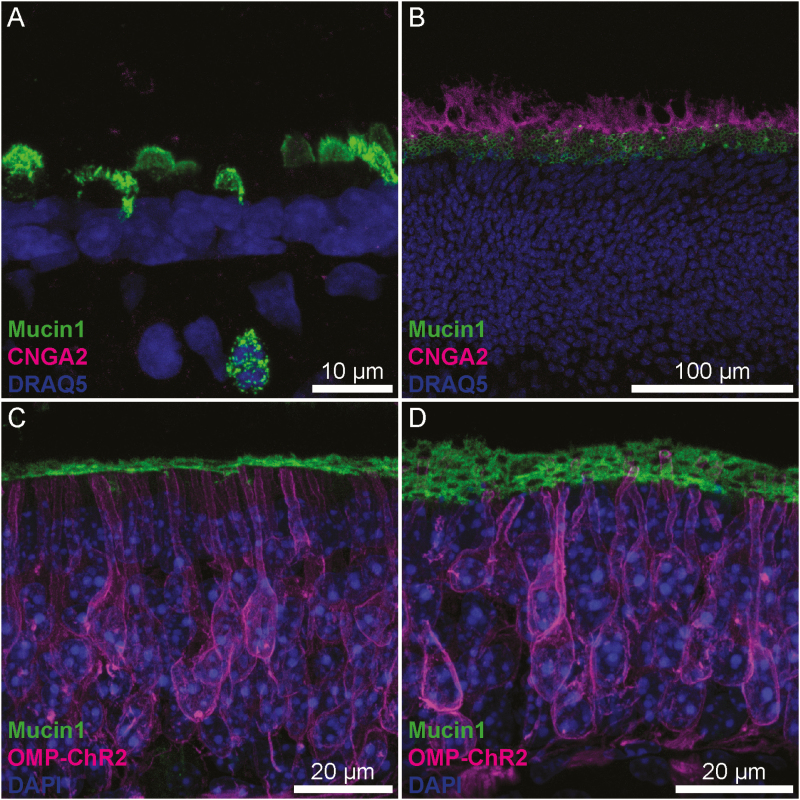
Figure 3.
Mucin 1 differential staining between OE and RE. (A) Mucin 1 is present in the apical layer of RE and expressed by scattered individual cells. (B and C) In the OE, mucin 1 lies at the base of the neurocilia. When viewed in oblique section (B), mucin 1 exhibits a lattice-like staining pattern indicative of perforations by OSN dendritic knobs suggesting it may be produced and secreted into this layer by the sustentacular cells. (C and D) OMP-ChR2-YFP knock-in mice were used to examine the relationship between OSNs (magenta) and mucin 1 (green). Mucin 1 is found in a layer just above the sustentacular cells, at the level of the OSN dendritic knob, as olfactory dendrites coursing through the OE are seen. In orthogonal section, this layer appears continuous, but in an oblique section (D) the lattice pattern of mucin 1 is clearly observed.

