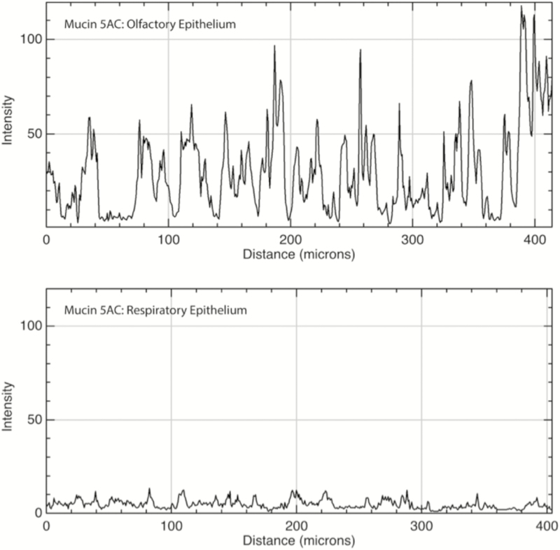
Figure 5.
Immunofluorescence quantitation of mucin 5AC in OE and RE. Top: Mucin 5AC was present primarily in submucosal glands within the OE and to a much lesser degree within the surrounding lamina propria connective tissue. Bottom: In the RE, there was a low level of immunofluorescence within the connective tissue and very few glands expressing the protein (none are present in the sample mentioned earlier). X axis is distance along a linear region of interest consisting of the submucosa, parallel to the epithelium; Y axis is intensity of immunofluorescence with possible values ranging from 0 to 255.

