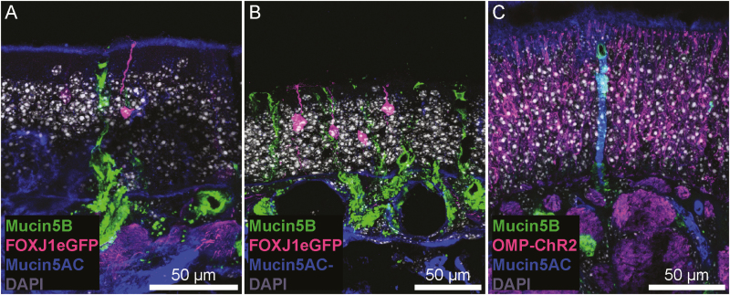
Figure 6.
Mucin 5AC and 5B staining in OE. (A and B) FOXJ1-eGFP mice were used to visualize a small subset of OSNs. The cilia and dendritic knob of an OSN (magenta) are observed within a layer of mucin 5AC immunoreactivity (blue; A), though this layer was not consistent across the entire OE (B). Additionally, mucin 5B+ (green), AC—Bowman’s glands are present in the OE. (C) Some Bowman’s glands were immunoreactive for mucin 5AC, surrounded by OMP-ChR2+ OSNs.

