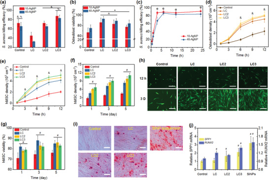
Figure 4.

a) S. aureus killing efficacy and b) osteoblast viability of various microcages with AgNP loading and the control (50 × 10−6 m AgNP suspensions). c) S. aureus killing kinetics of LC3 microcages loaded with AgNPs. Cell density of d) osteoblast cells and e) hMSCs on control substrate and substrates coated with microcages incorporated with SiNPs within 12 h. f) Proliferation of hMSC and g) percentage of live cells on various substrates at 1, 3, and 5 days. h) CLSM images of hMSC adhesion at 12 h and 3 days on samples in (e)–(g). Scale bars in (h) are 200 µm. i) Mineralized extracellular matrix production and j) evaluation of osteogenic potential of hMSCs cultured on various nanocoatings. Scale bars in (i) are 50 µm. The mineralized extracellular matrix was stained with Alizarin Red S at day 14. Glyceraldehyde‐3‐phosphate‐dehydrogenase (GAPDH) was carried out as an endogenous control. & p < 0.01 compared to LC microcages. % p < 0.05 compared to LC2 microcages. * p < 0.01 compared to the control. ^ p < 0.05 compared to LC3 microcages. @ p < 0.01 compared to 0.5 h. # p < 0.01 compared to the controls.
