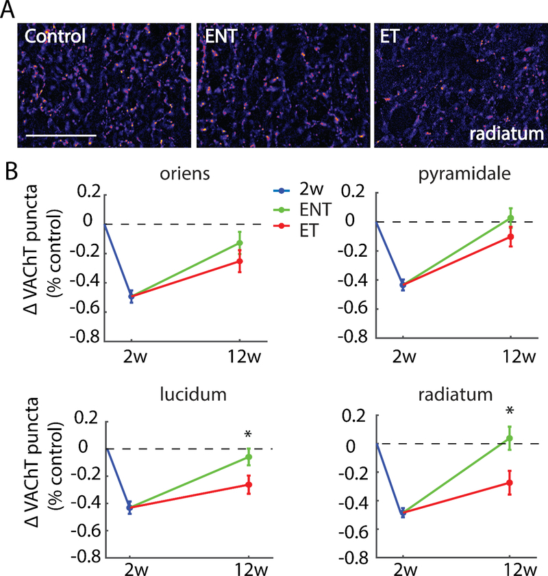
Figure 5. Tinnitus animals exhibit persistent decreases in VAChT labeling relative to no-tinnitus animals in synapse-rich areas of hippocampal area CA3.
(A) Representative images from stratum radiatum at 400x magnification; Scale bar = 50 μm. (B) Mean (±SEM) change of VAChT density (normalized to respective control) in area CA3 in two-weeks and 12-weeks post-noise-exposure animals. Strata lucidum (s.l) and radiatum (s.r) showed significantly lower VAChT labeling in tinnitus animasl (ET) than in no-tinnitus animals (ENT), whereas strata oriens (s.o) and pyramidale (s.p) showed similar VAChT labeling in ENT and ET animals; *p < 0.05.

