. 2020 Mar 30;40(1):72–88. doi: 10.7705/biomedica.4762

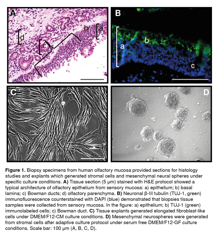
Figure 1. Biopsy specimens from human olfactory mucosa provided sections for histology studies and explants which generated stromal cells and mesenchymal neural spheres under specific culture conditions. A) Tissue section (5 µm) stained with H&E protocol showed a typical architecture of olfactory epithelium from sensory mucosa: a) epithelium; b) basal lamina; c) Bowman ducts; d) olfactory parenchyma. B) Neuronal (3-III tubulin (TUJ-1, green) immunofluorescence counterstained with DAPI (blue) demonstrated that biopsies tissue samples were collected from sensory mucosa. In the figure: a) epithelium; b) TUJ-1 (green) immunolabeled cells; c) Bowman duct. C) Tissue explants generated elongated fibroblast-like cells under DMEM/F12-CM culture conditions. D) Mesenchymal neurospheres were generated from stromal cells after adaptive culture protocol under serum free DMEM/F12-GF culture conditions. Scale bar: 100 µm (A, B, C, D).

Figure 1