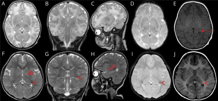
Figure 3. MRI of the index patient (II:3).
Images at ages 23 days (A–E) and 4 years (F–J). T2-weighted MRI in axial (A and F), coronal (B and G), and sagittal plane (C and H), axial GRE imaging (D and I), and T1-weighted imaging in axial plane (E and J). At age 4 years, a persistent high signal is perceived in the basal part of the bilateral globus pallidi (Δ) (F), which is not seen at age 23 days (A). The caudate part of the caudate nuclei (arrow head) presents low signal on T2 in all 3 planes (F–H) including GRE imaging (I) with high signal on T1 (J) compared with normal appearance at age 23 days (A–E). Only the normal mature lateral geniculate body of the thalamus (arrow) is seen at age 23 days just medially to the caudate part of the caudate nucleus (E). GRE = gradient echo sequences.

