FIG 2.
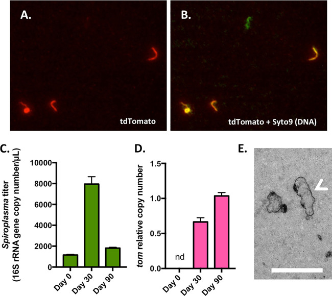
Detection of S. poulsonii transformant by microscopy and molecular methods. (A) Representative image of S. poulsonii transformants producing the red fluorescent protein tdTomato. (B) Overlay of panel A with a Syto9 signal (DNA staining) showing all bacteria. A fluorescent-green-only, probable spontaneously tetracycline-resistant bacterium is visible in the center. (C) Absolute quantification of S. poulsonii 16S rRNA gene copy number in the transformant culture. (D) Quantification of the tdTomato gene copy number relative to the 16S rRNA gene copy number. nd, not detected. (E) Transmission electron microscopy image of BSK-H-spiro solid medium showing the transformant cells scattered inside the agar gel. The white arrow indicates a cell that has been cut longitudinally through the agar matrix, revealing the characteristic helical morphology of Spiroplasma. Scale bar, 500 nm.

