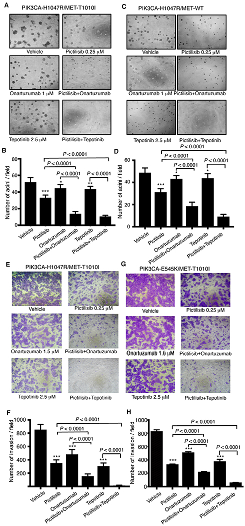
Figure 4. Effects of combined targeting of PI3K and MET on cell growth in Matrigel or invasion in MCF-10A derived cells.

A total of 4 × 10³ MCF-10A–derived cells expressing PIK3CA-H1047R/MET-T1010I (A) or PIK3CA-H1047R/MET-WT genes (C) were resuspended in modified growth medium (2.5% horse serum and 5 ng/ml EGF) supplemented with HGF (40 ng/ml) and 2% growth factor reduced Matrigel and treated with various drugs as indicated. The medium was exchanged every 3 days. Photographs (×40) of representative fields were taken on day 7. The figures shown are mean ± standard deviation of triplicates, representative of two independent experiments (*, P < 0.01; **, P < 0.001; ***, P < 0.0001 vs. vehicle, ANOVA) (B, D). The MCF-10A–derived cells expressing PIK3CA-H1047R/MET-T1010I (E) or PIK3CA-E545K/MET-T1010I (G) were starved for 20 hours in serum-free DMEM-F12 lacking EGF, insulin, and hydrocortisone. A total of 1 × 10⁵ cells were inoculated into the upper chamber. pictilisib, onartuzumab, or tepotinib alone or in combinations as indicated were added into both the upper and lower chambers. HGF and fibronectin were added in the lower chamber as the inducer. Invasive cells were photographed and counted in 10 random fields. The figures shown are mean ± standard deviation of triplicates, representative of two independent experiments (***, P < 0.0001 vs. vehicle, ANOVA) (F, H).
