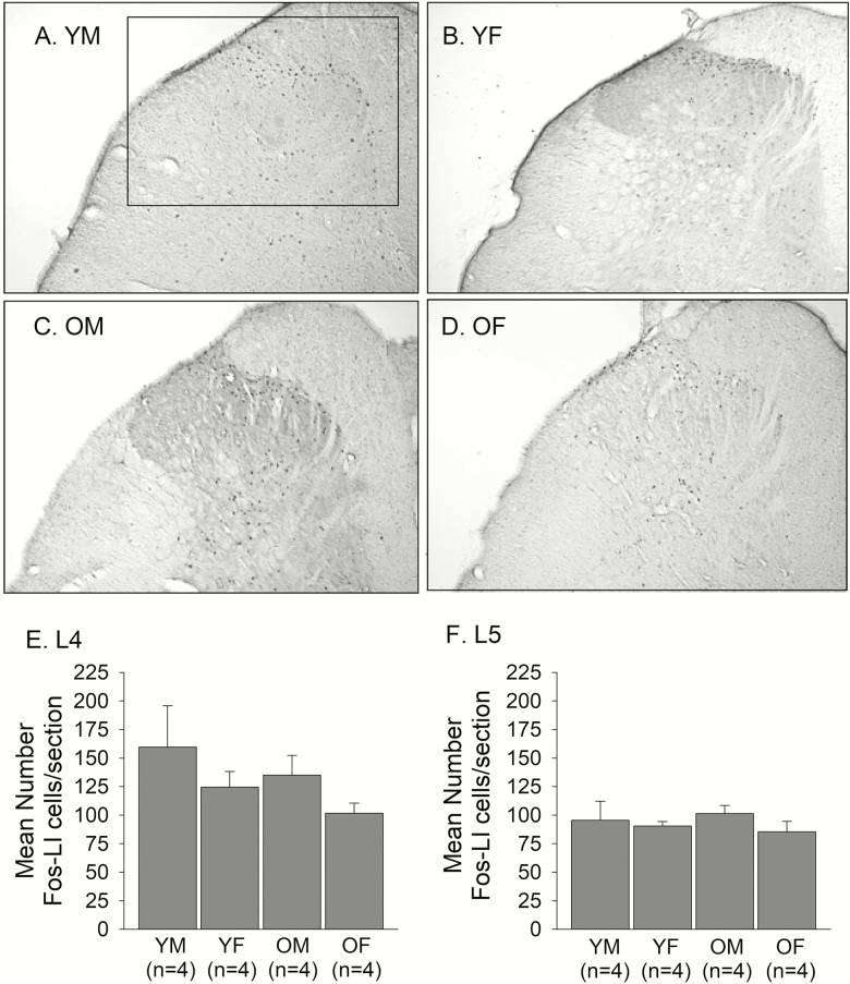
Figure 3.
Representative photomicrographs showing capsaicin-induced Fos-LI neurons in the dorsal horn of the L4 spinal cord of (A) YM, (B) YF, (C) OM, and (D) OF. The box shown in A indicates the area in the upper quadrant of the dorsal horn where all counts of Fos-LI were made. The same box was applied across all sections in all experimental groups. All counts were made from photomicrographs taken at 10 x magnification. The average number of capsaicin-induced Fos-LI neurons per section in the dorsal horn of the (E) L4 and (F) L5 spinal cord were compared between YM, YF, OM, and OF.

