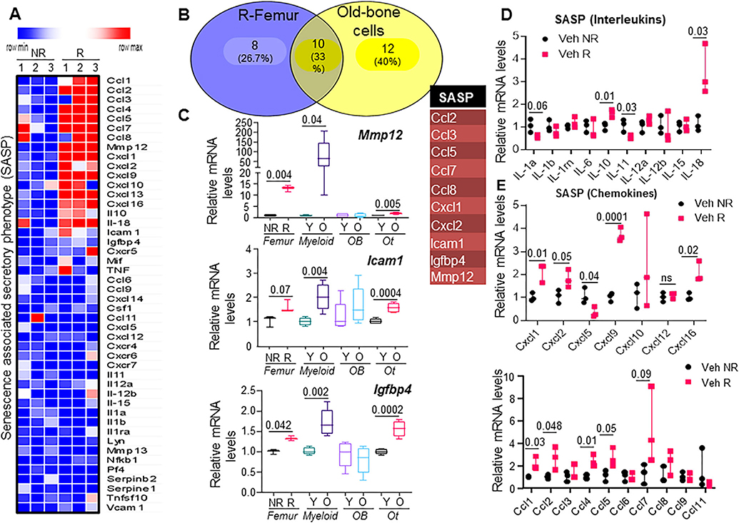
Fig. 3.
Radiation of bone induces a SASP similar to that of aged bone cells. (A) Heat map of SASP genes from NR and R femurs (n = 3 mice) at 14 days post-FRT. (B) Venn diagram representing SASP genes that were commonly expressed between R femurs from 4-month-old male mice and old bone cells from 24-month old male mice (table). (C) Comparison of expression levels of SASP genes Mmp12, Icam1, and Igfbp4, between R bones and NR bones at 14 days post-FRT, with enriched cell populations from bones of young (Y) versus old (O) animals. y axis: fold change normalized against NR (for comparison between NR versus R), and normalized against Y (for comparison between Y versus O). Expression of SASP genes that are classified as interleukins and chemokines are shown (D and E, respectively). y axis: fold change normalized against NR (for comparison between NR versus R). Statistical analysis was done using GraphPad Prism and p value was calculated using a two-tailed paired t test to compare the R and NR bones from the same animals, and using a two-sampled t test for comparison of young and old bone cells.

