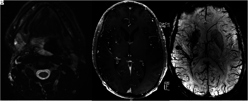
FIG 4.
A 38-year-old man with extensive airway malformation. A, T2 FS MR imaging shows an extensive venous malformation involving the airway and tongue base. B, Axial postcontrast MR imaging shows extensive DVAs of the right frontal lobe, basal ganglia, and temporal lobe. C, 7T SWI shows the extensive DVAs and multiple cavernomas in the radicles of the DVAs.

