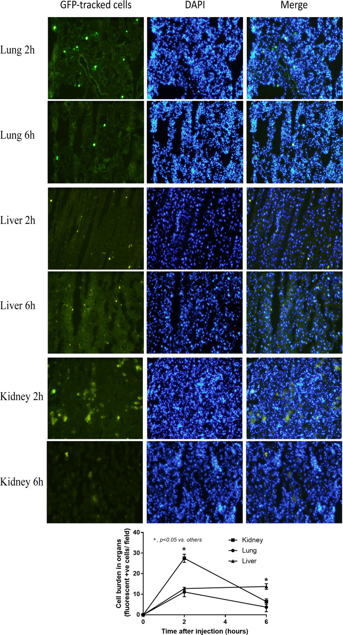
Fig 6. Macrophages are demonstrated in kidney and liver at 2 h and 6 h after administration.
The representative pictures of the kinetics of tagged green fluorescent protein (GFP) macrophages after intravenous injection in different organs at different time-points and the analysis of cells burdens in organs are demonstrated. DAPI, 4′, 6-diamidino-2-phenylindole (a DNA fluorescence staining color).

