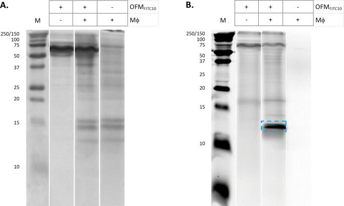
Fig 3. Tris-glycine SDS-PAGE of conditioned media.
Conditioned media from cultures of OFMFTIC10, OFMFITC10+Mϕ, and Mϕ alone were separated by Tris-glycine SDS-PAGE electrophoresis. Tris-glycine gels were stained with either Coomassie (A) or imaged via a fluorescence scanner (B). The ~12 kDa protein band of interest highlighted in panel B (blue). Unedited gel images are provided in Supporting Information (S2. File).

