
Figure 2.
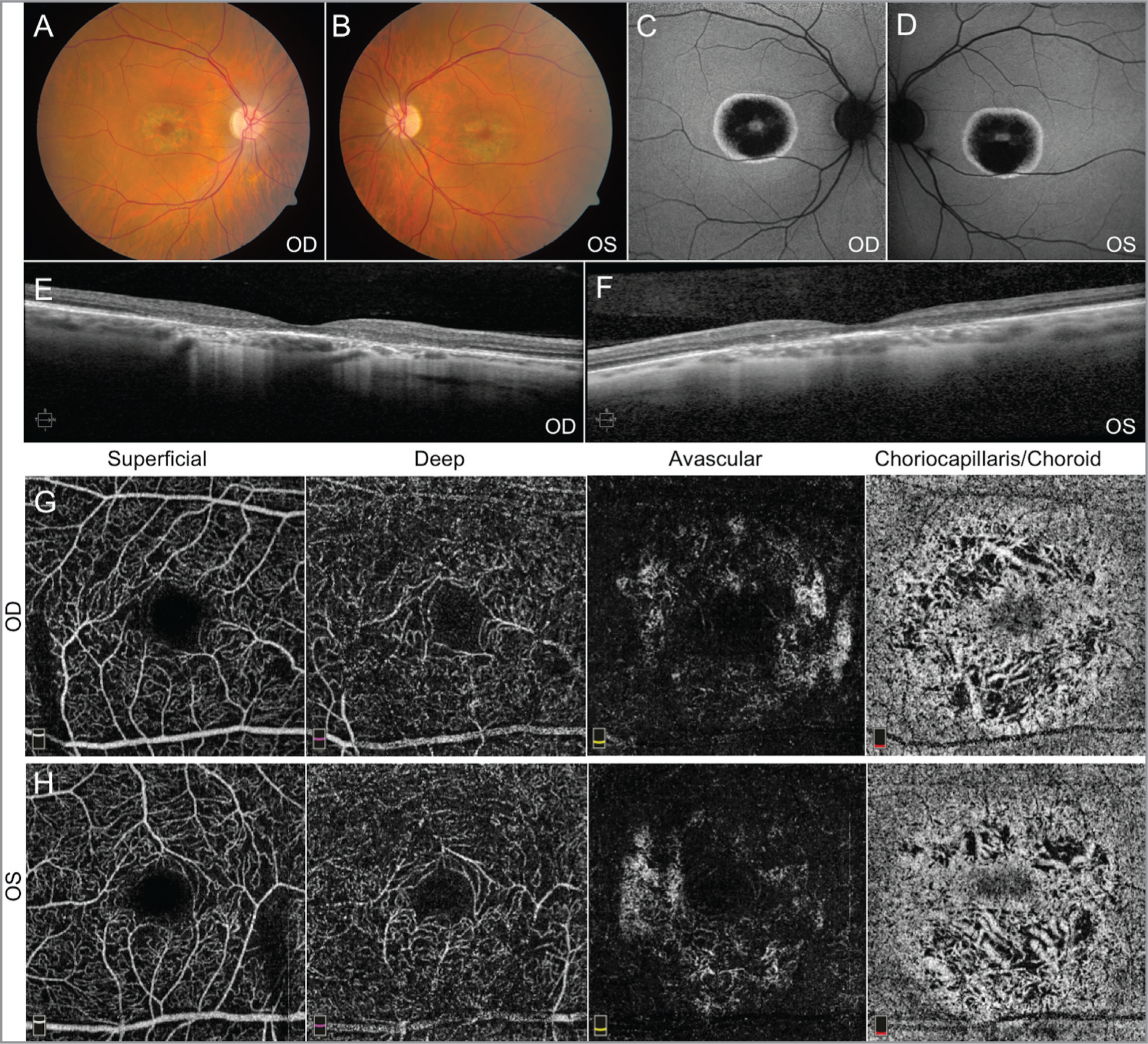
Fundus imaging from Patient 2. Fundus photos (A, B) and autofluorescence (C, D) demonstrate pigmentary changes with bull’s-eye maculopathy. Spectral-domain optical coherence tomography (OCT) of the macula (E, F) is consistent with outer retinal degeneration. OCT angiography imaging (G, H) showed moderate attenuation of vasculature in the choriocapillaris/choroid layer. These findings appear more pronounced than those found in Patient 1.