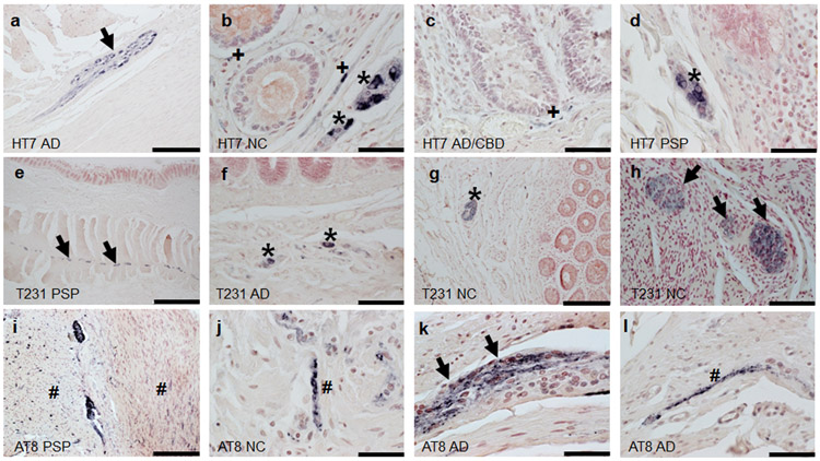
Figure 5.
Phosphorylated tau immunoreactivity within the sigmoid colon. Diagnostic group and antibody used are listed in the lower left corner of each photo. Immunoreactivity of the myenteric plexus is indicated by black arrows, muscularis by #, mucosa by +, and submucosa as *. b-d; j-l scale bar=50um a, g, i scale bar=200um; e scale bar=1 mm f,h scale bar=100um.

