Fig. 1.
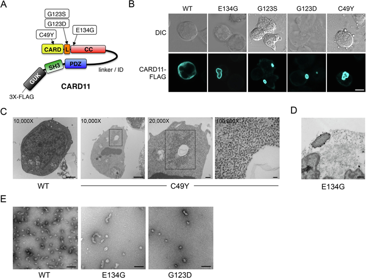
Gain-of-function CARD11 mutants form mCADS. (A) Cartoon of CARD11 protein indicating approximate locations of BENTA-associated GOF mutations described. L = LATCH domain, CC = coiled-coil. (B) Confocal immunofluorescent (IF) microscopy analysis of BJAB B cells transfected with WT and GOF CARD11-FLAG mutants, detected using anti-FLAG-AlexaFluor 647 Ab. Merge image includes DAPI staining. Scale bar = 5 μm. (C) JPM50.6 T cells were transfected with WT or C49Y CARD11 and fixed for TEM analysis 24 hrs post-transfection. Numbers indicate magnification, squares denote putative mCADS that are not found in WT cells (n = 30 cells analyzed for each). Data are representative of two independent experiments performed with WT, C49Y, E134G, or G123D CARD11. (D) ImmunoGold staining and TEM analysis of JPM50.6 T cells transfected with E134G CARD11 as prepared in Supp. Fig. 2A. Data are representative of 2 independent experiments. (E) TEM micrographs of recombinant CARD11 (aa 8–302) showing heterogeneous clusters of WT (20–40 nm), E134G (60–120 nm), and G123D (20–40 nm).

