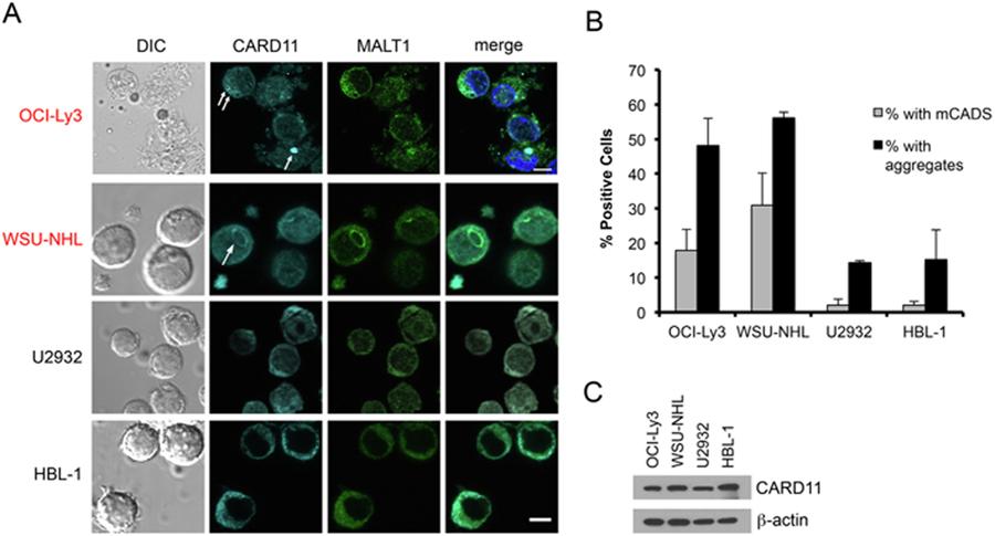
Fig. 7.
Endogenous mCADS formation in DLBCL lines carrying GOF CARD11 mutations. (A) Confocal IF microscopy of DLBCL cell lines stained with anti-CARD11 and anti-MALT1 Abs. Lines marked in red carry GOF CARD11 mutations. Merge images include DAPI staining. Scale bar = 5 μm. (B) The % of cells with mCADS (gray bars) and/or visible aggregates (black bars) was scored in multiple fields (>100 cells/DLBCL line). Average ± SD was calculated from 2 independent scorers. (C) Immunoblot analysis for relative CARD11 protein expression in lysates prepared from DLBCL lines. All data are representative of >3 independent experiments.

