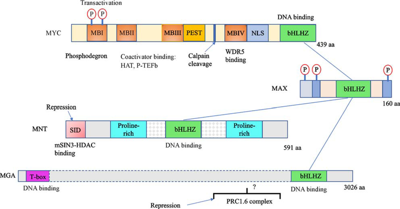
Fig. 3.
Organization of the MYC, MAX, MNT and MGA proteins. Heterodimers are formed by direct interaction of the basic-helix-loop-helix-zipper (bHLHZ) domain of MAX with the bHLH-Z domains of either MYC, MNTor MGA (blue lines). Number of residues in each protein indicated at C terminus. MYC: MBI–IV — conserved MYC boxes; PEST— region rich in proline, glutamic acid, serine and threonine; NLS— nuclear localization sequence; Calpain cleavage site—proteolytic cleavage to generate MYC-Nick [2]. MNT: SID — binding site for the mSIN3 co-repressor complex. MGA: repression mediated through assembly into a variant polycomb repressor complex (PRC1.6). Question mark indicates that the region of MGA that directly interacts with the complex is unknown. Protein lengths not to scale. See text for details.

