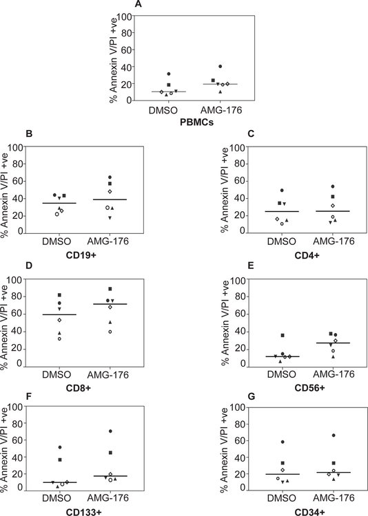
Fig 3. Biological impact of AMG-176 in normal PBMCs.
Normal PBMCs were isolated from peripheral blood sample of healthy donors. PBMCs were cultured in media supplemented with 10% human serum. Each sample was treated with either dimethyl sulfoxide (DMSO) (0.1%) or 300 nM AMG-176 for 24 hours. Cell death was determined using flow cytometry after Annexin V/propidium iodide (PI) staining and cell surface marker staining. A. PBMCs, B. CD19+ B-cells. C. CD4+ T-cells. D. CD8+ T-cells. E. CD56+ NK-cells. F. CD133+ plasma cells and G. CD34+ myeloid cells.

