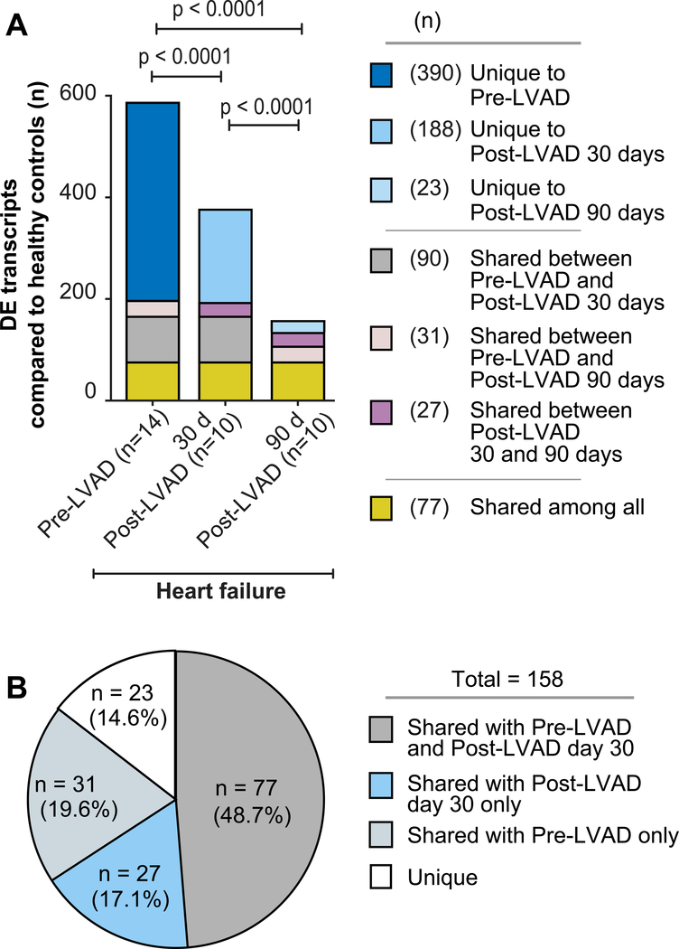
Figure 4. The expression of many transcripts in platelets normalizes in heart failure patients following LVAD implantation.
(A) Comparison of the number of significantly differentially expressed (DE) transcripts (FDR<0.05) in platelets from heart failure patients followed longitudinally over time (e.g. prior to LVAD implantation (n=14), 30 days (n=10), and again 90 days (n=10) following LVAD implantation), as compared to matched healthy controls (n=7). As denoted in the legend to the right, “Unique” refers to transcripts in platelets that were significantly differentially expressed in heart failure patients, compared to controls, at just one time point. “Shared” refers to transcripts in platelets that were durably and significantly differentially expressed in heart failure patients, compared to controls, at two or more time points. (B) Pie chart showing the distribution of significantly differentially expressed transcripts in platelets from heart failure patients 90 days following LVAD implantation, as compared to platelet transcriptomic assessments 30 days following LVAD implantation (Post-LVAD day 30) or prior to LVAD implantation (pre-LVAD). “Unique” and “Shared” denote the same as in panel A.

