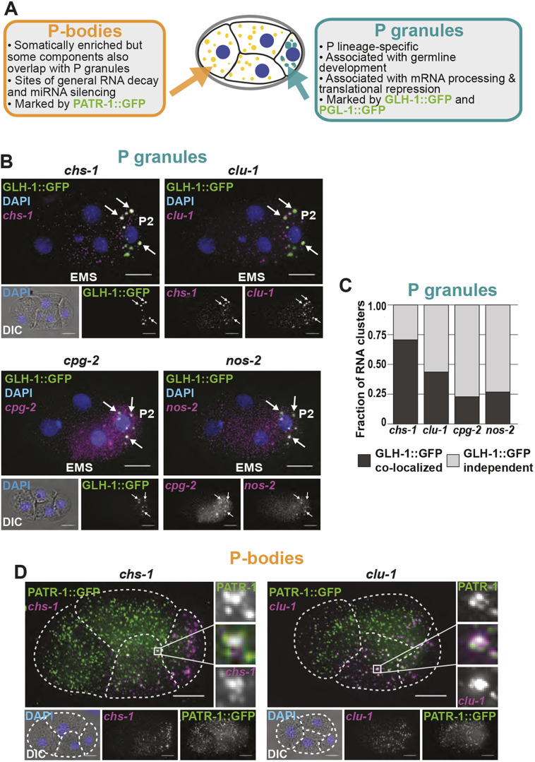
Fig. 3.
Posterior clustered mRNAs co-localize with P granules and P-bodies. (A) Schematic detailing how P granules are distinct from P-bodies. (B) Fixed embryos were imaged for the P granule marker GLH-1::GFP (green) and chs-1, clu-1, cpg-2 or nos-2 transcripts (magenta). DNA (DAPI, blue) and differential interference contrast microscopy (DIC) are also shown. (C) The fraction of mRNA clusters overlapping with P granules (dark gray) and P granule-independent clusters (light gray) in four-cell embryos was calculated by assessing spatial overlap between mRNA clusters and GLH-1::GFP-marked P granules. (D) Fixed embryos were imaged for the P-body protein marker PATR-1::GFP amplified using immunofluorescence (green) with smFISH imaging of chs-1 mRNA or clu-1 mRNA (magenta), and DNA (DAPI; blue). Enlargements of boxed areas illustrate regions of co-localization. Dashed white lines indicate cell boundaries.

