Figure 2.
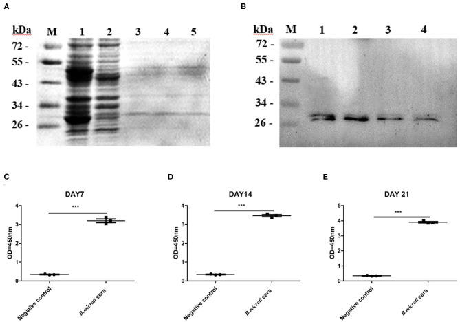
Recombinant antigen production and evaluation of rBmSP44 as a diagnostic antigen. (A) SDS-PAGE analysis of bacterial lysate as stained by Coomassie blue. Lane 1: induced protein; Lane 2: non-induced control; Lane 3 and 5: The purification of rBmSP44 after cleavage of GST tag; (B) Western blot analysis of rBmSP44; M: Protein marker; Lane 1–4: The recombinant proteins; Protein incubated with an anti-rBmSP44 monoclonal antibody (~24 kDa); (C–E) rBmSP44 recognized by sera from B. microti infected mice [Sera from day7 (C), 14 (D), and 21 (E) p.i.] Significant differences are as follows: ***P < 0.001, compared to non-infected control sera, t-test. The data shown are representative of at least 3 independent experiments.

