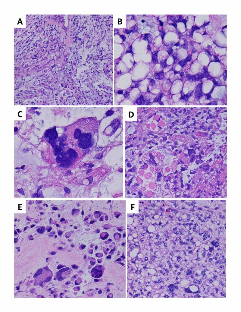
Figure 1. Histologic sections demonstrate a pleomorphic liposarcoma, a high-grade malignancy with aggressive biologic behavior, and morphologic features overlapping with other pleomorphic mesenchymal lesions and liposarcomas.
(A) The initial core needle biopsy showed areas with a myxoid background, scattered atypical tumor cells, and focal curvilinear vessels, resembling an intermediate grade myxofibrosarcoma, a pattern well-documented to occur in a significant proportion of pleomorphic liposarcomas (H&E stain, 100x magnification); (B) the initial core needle biopsy also demonstrated extensive adipocytic differentiation, which can occur in pleomorphic liposarcomas and obscure the main diagnostic feature, i.e., characteristic lipoblasts with vacuoles crisply indenting the nuclear contours for a “scalloped” appearance. Lipoblasts are required for the diagnosis of pleomorphic liposarcoma (in contrast to other liposarcoma subtypes in which they are neither necessary nor sufficient). Lipoblasts can be highly focal and variable in extent, necessitating very thorough tissue sampling and representing a well-known pitfall in misdiagnosis, particularly in small biopsies such as this case (H&E stain, 1000x magnification); (C) extreme pleomorphism and bizarre atypia is a common finding, in contrast to myxoid/round cell liposarcoma (H&E stain, 1000x magnification); (D) intracytoplasmic eosinophilic globules are reported in association with pleomorphic liposarcoma (H&E stain, 400x magnification); (E) the surgical resection demonstrated widespread viable tumor with florid pleomorphism, reflecting neoadjuvant therapy-related effects, and precluding definitive subtyping beyond an unclassifiable pleomorphic sarcoma in the absence of other diagnostic samples from different time points (H&E stain, 1000x magnification); (F) a biopsy of the liver metastasis again showed hypercellularity, prominent adipocytic differentiation, and marked pleomorphism (H&E magnification, 400x magnification)
H&E: hematoxylin & eosin

