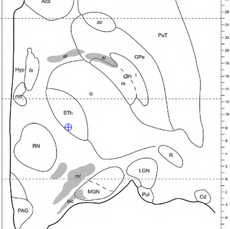
Figure 2. Mapping a 3D intraoperative coordinate to a point on the closest atlas section from a series. Here, we use the current point illustrated in Table 4, {-4.4, -10.3, -4.2}. Using the method, the closest axial slice of the Morel Atlas maps to {-4.4, -10.3, -4.5}. The position of the electrode tip is observed within the margins of the subthalamic nucleus (STN).
All Coordinates are given as {AP, LAT, VERT}
3D = Three Dimensional
Morel Atlas: please refer to reference [17].

