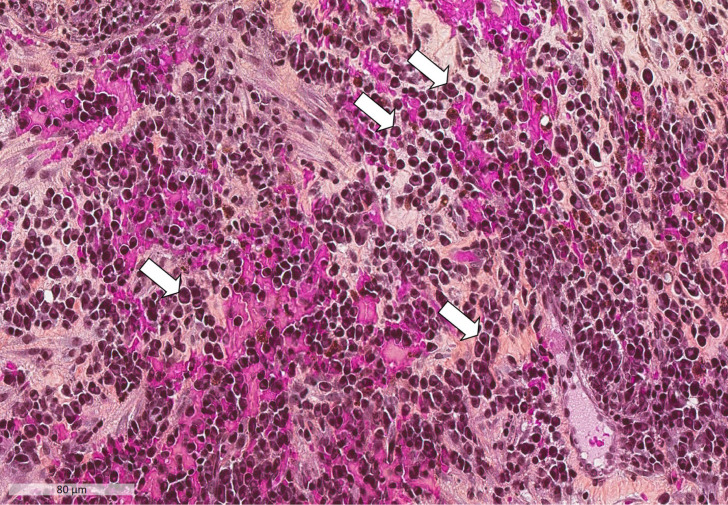
Figure 1.
Plasma cell infiltration into synovial tissue (Hematoxylin-Eosin-Safran; x200 magnification). Numerous plasma cells are present. The white arrows indicate some of the plasma cells: they consist of an eccentric nucleus with heterochromatin in a characteristic cartwheel or clock face arrangement, surrounded by an abundant basophilic cytoplasm.

