Figure 3.
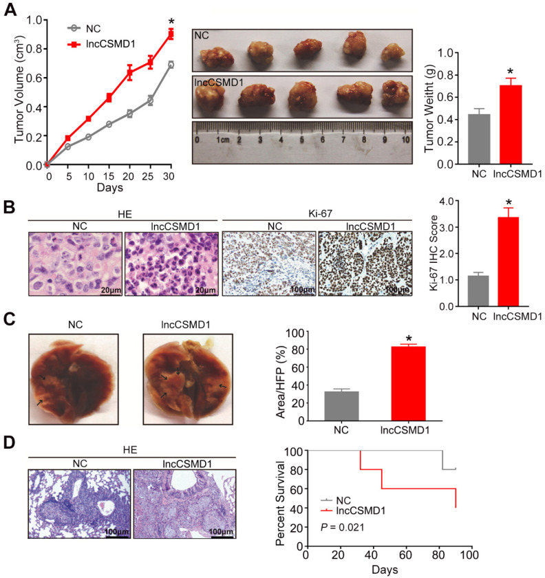
LncCSMD1 promotes growth and metastasis of xenograft tumors derived from Hep3B HCC cells. (A) In the tumor growth curve, the tumors grow faster in the mice subcutaneously inoculated with Hep3B cells overexpressing lncCSMD1 than in mice with the HCC cells carrying a control vector (left); Images of xenograft tumors derived from Hep3B cells stably overexpressing lncCSMD1 or carrying a control vector (middle); the weights of xenograft tumors in the two groups were compared with histogram (right), in which data were analyzed by sample-paired t test; *P <0.05 (the followings are the same). (B) Representative images of HE staining (left) and Ki67 IHC staining (middle) displayed the pathological configuration and proliferation capacity of HCC xenograft cells; IHC scores for Ki67 were compared in the two groups with histogram (right). (C) Representative images of nude mouse lungs with metastatic foci derived from tail-vein injected Hep3B cells stably overexpressing lncCSMD1 or carrying a control vector (n=5 mice/each group); the metastatic foci ratios (foci to the observed lung area) were compared in the two groups with histogram. (D) Representative images of metastatic foci stained with HE in the lung tissue sections (left); survivals mice tail-vein injected with Hep3B cells stably expressing lncCSMD1 or a control vector were analyzed with Kaplan-Meier curve (right).

