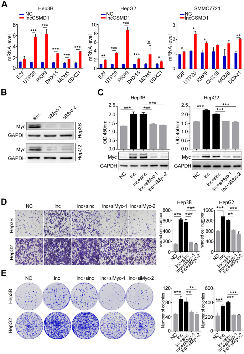
Figure 6.
MYC protein plays a key role in lncCSMD1-induced malignant phenotypes of HCC cells. (A) MYC target genes in Hep3B, HepG2 and SMMC7721 cells with overexpression of lncCSMD1 are expressed higher than those in the control cells, as shown by qRT-PCR. (B) MYC protein expression is reduced by siRNA against MYC in Hep3B and HepG2 cells, as shown by Western Blot. (C) MYC protein in Hep3B and HepG2 cells with lncCSMD1 overexpression is elevated, as displayed by western blot (lower panels) and promotes cell proliferation as shown by CCK-8 assay (upper panels) compared with that in control cells; when the HCC cells were treated with siMYC again, MYC protein was reduced and leads to decrease in cell proliferation. (D, E) The same treatments as in the above (C) were conducted in the same Hep3B and HepG2 cells, and these cells were determined with transwell assay (D) and colony formation assay (E), in which overexpressed lncCSMD1 promotes the invasion and colony formation of the HCC cells and the downregulated MYC by siRNA inhibits colony formation and invasion of the HCC cells.

