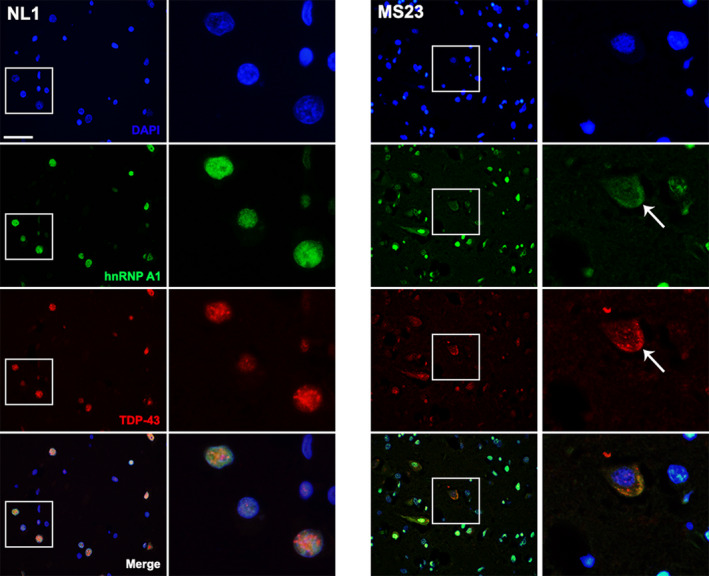
Figure 4.

Mislocalization of hnRNP A1 and TDP‐43 within the same neurons in MS normal appearing cortex. Immunofluorescence shows colocalization of hnRNP A1 and TDP‐43 in the cytoplasm of neurons from MS brain. Control brain (NL1) shows nuclear (DAPI‐blue) localization of hnRNP A1 (green) and TDP‐43 (red) and colocalization in the nucleus (merged image). A representative neuron from MS (MS23) shows that there is decreased nuclear staining and cytoplasmic accumulation of hnRNP A1 and TDP‐43 (arrow) suggesting colocalization of mislocalized hnRNP A1 and TDP‐43 within the same neuron. Scale bar 50μm. hnRNP A1, heterogeneous nuclear ribonucleoprotein A1; TDP‐43, TAR‐DNA‐binding protein‐43; MS, multiple sclerosis.
