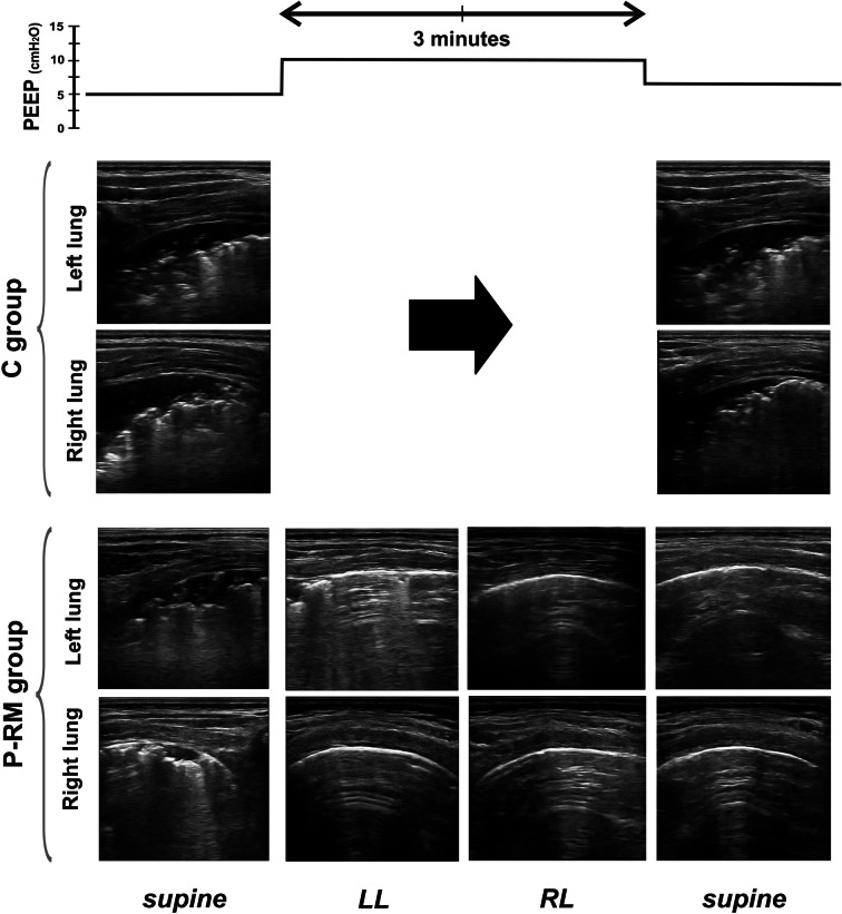
Fig. 4.
Example of the protocol in one representative patient per group. LUS images were assessed in the posterior areas in supine (dorsal lung) and in the uppermost areas in the lateral positions (ventral lung). C group control patients, P-RM postural recruitment patients, LL left lateral, RL right lateral position. Note typical atelectasis with air bronchograms before the 10-PEEP maneuver in both groups and how only the P-RM resolved it

