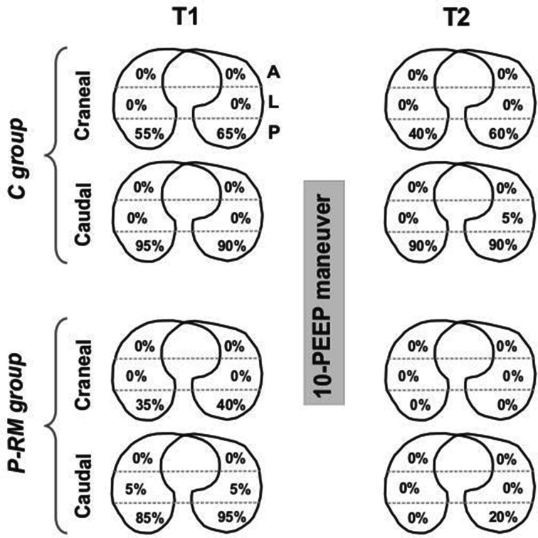
Fig. 5.
Distribution of atelectasis between groups along the study. C group control patients, P-RM postural recruitment patients, A anterior, L lateral, P posterior lung zones assessed by lung ultrasound. Cranial = axial cut representing the superior thoracic area above a horizontal line crossing the nipples. Caudal = axial cut representing the inferior thoracic area below a horizontal line crossing the nipples. % = the percent of all patients per group that presented atelectasis in a particular lung zone

