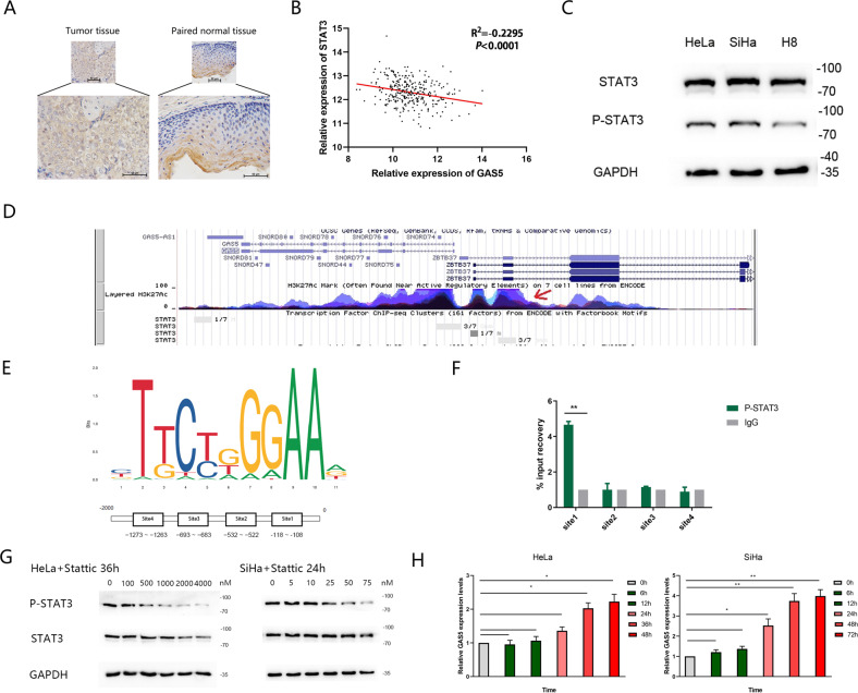
Fig. 2. P-STAT3 binds with the promoter of GAS5 and modulates its transcriptional level.
a Immunohistochemical staining for P-STAT3 in cervical cancer tissues and adjacent normal tissues (magnification, ×200). Scale bar, 50 μm. b The correlation between STAT3 and GAS5 expression in CC tissues from the TCGA database was analysed by Pearson correlation analysis. c Western blot analysis showed that the expression levels of P-STAT3 in CC cells were higher than those in normal cervical cell, whereas the total STAT3 level did not differ. d UCSC database analysis revealed that the promoter of GAS5 had high enrichment of STAT3. e The binding motif of the transcription factor STAT3 was predicted via the JASPAR CORE database. f A ChIP assay demonstrated P-STAT3 binding to site 1 (−118 to −108) in the GAS5 promoter region. **P < 0.01 (Student’s t-test). g Western blot analysis showed that the P-STAT3 level changed dose-dependently under treatment with Stattic, whereas the total STAT3 level remained unchanged at Stattic concentrations of 0–50 nM for SiHa cells and 0–1000 nM for HeLa cells. GAPDH was used as the control. h A decline in the P-STAT3 level was accompanied by an increase in the GAS5 level. Total RNA was isolated from SiHa cells treated with 50 nM Stattic and HeLa cells treated with 1000 nM Stattic for different durations. GAS5 levels were examined by qRT-PCR. β-Actin was used as the input control. The fold change was calculated with respect to the control by the 2−ΔΔCt method. The data are shown as the means ± SDs (n = 3). *P value <0.05 compared to untreated cells.

