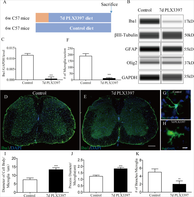
Fig. 1. Efficient depletion of microglia in the adult spinal cord via pharmacologic inhibition of CSF1R.
a Schematic of the experimental design: 6-week-old C57BL/6 mice were fed either PLX3397 or control chow for 7 days. On day 7, mice were euthanized for western blots or Immunostaining. b Western blot analysis of spinal cord homogenates for steady state levels of the microglia marker Iba1, neuronal markers βIII Tubulin, the oligodendrocyte marker Olig2, and the astrocyte markers GFAP. c Quantification of Iba1 in (B) showing CSF1R inhibition decreased Iba1 protein levels (n = 4 per group). d, e Representative immunofluorescence images of mouse spinal cord sections showing Iba1+ ramified microglia (green). Almost no Iba1+ cells were detected in mice that were fed a PLX3397 diet for 7d. f Quantification of the number of IBA1+ cell in the spinal cord from control and PLX3397-treated mice (n = 4 per group) as shown in (D) and (E). g, h Iba1 immunostaining shows changes in microglia morphology with representative microglia shown from control and 7-day PLX3397-treated mice. I-K Microglial morphology were assessed by the diameter of cell body per microglia (i), the process diameter per microglia (j) and the number of branches per microglia (k) (n = 4 per group). Data are expressed as mean ± SD. Scale bars: d, e, in e 200 µm; g, h, in h 10 µm.

