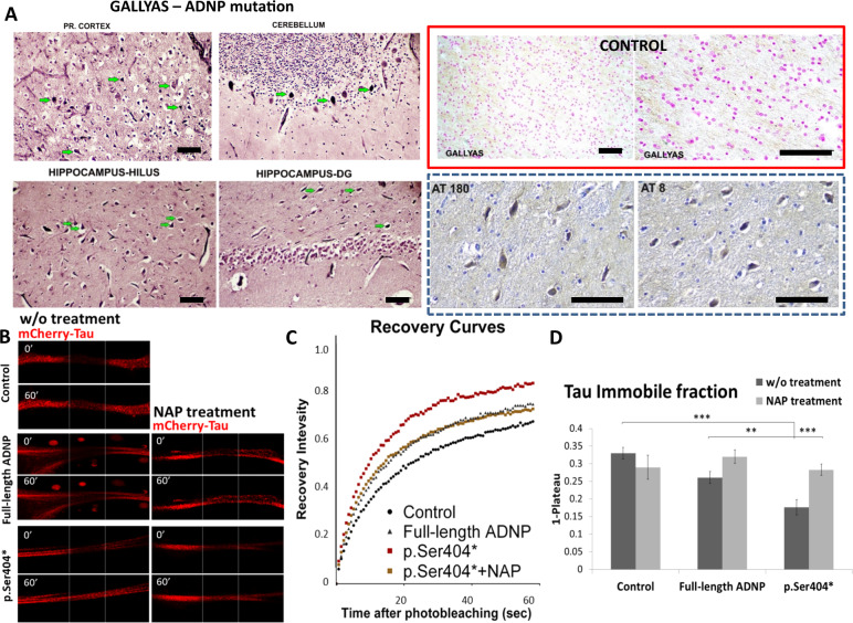
Fig. 1. Discovery of ADNP syndrome juvenitle postmortem tauopathy and potential rescue by NAP treatment.
a The ADNP syndrome juvenile brain exhibits intensive tauopathy. Gallyas staining (Tau pathology) is indicated by green arrows. Frontal cortex (Pr cortex), cerebellum, hippocampal hilus, and dentate gyrus showed positive staining. In the hippocampus the positive cells were in the hillus and in the molecular area, in the granular area of dentate gyrus, CA1, CA2, CA3, there were no positive cells. While no apparent Gallyas staining was observed in the control hippocampus (red box), the ADNP case study tauopathy was verified by hippocampal hyperphosphorylated Tau immunohistochemistry (AT180 and AT8 antibodies). Bars represent 100 μm. The ADNP snippet, drug candidate. b NAP enlists Tau to microtubules in the face of ADNP mutations: protection against Tauopathy. Representative images of photo-bleaching (0′) and fluorescence recovery (60′) of mCherry-tagged Tau in differentiated N1E-115 cells co-transfected with GFP-tagged full-length ADNP or truncated p.Ser404* ADNP form with or without NAP treatment (10−12M, 4h). Transfection with backbone plasmid (pEGFP-C1) expressing non-conjugated GFP, was performed as a control. c Fluorescence recovery after photobleaching (FRAP) recovery curves of normalized data (see “Online Resources 1”). d The graph represents averages (±SEM) of the fitted data (from three independent experiments) of immobile fractions. Normalized FRAP data were fitted with one-exponential functions (GraphPad Prism 6) and statistical analyses was done by Two Way ANOVA (SigmaPlot 11, with all pairwise multiple comparison procedures—Tukey Test). Statistical significance is presented by **P < 0.01, ***P < 0.001. Control n = 64; control+NAP n = 14; full-length ADNP n = 55; full-length ADNP+NAP n = 46; p.Ser404* n = 33; p.Ser404* + NAP n = 62.

