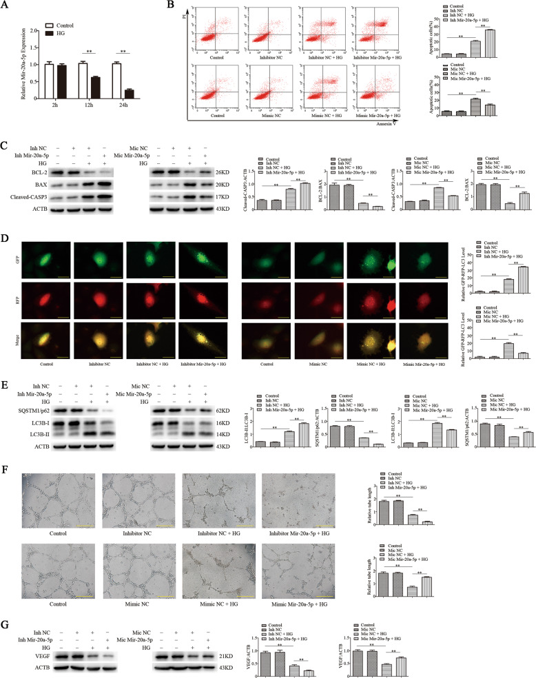
Fig. 1. Mir-20a-5p regulates apoptosis, autophagy, and angiogenic function of EPCs induced by high glucose in vitro.
EPC dysfunction was induced by high glucose (30 mM) treatment for 24 h. Mannitol was used as an osmolar control treatment. EPCs were transfected with mir-20a-5p or negative control (NC) inhibitor, or mir-20a-5p or NC mimic. a RT-qPCR analysis of mir-20a-5p expression. b Flow-cytometric analysis of apoptosis using annexin V/propidium iodide (PI). Annexin V+/PI+ or Annexin V+/PI− (quadrants 2 and 3) cells were defined as apoptotic cells. c Protein levels of BCL-2, BAX, and cleaved-CASP3 as detected by western blotting. d Representative images showing LC3 staining in different groups of EPCs infected with GFP-RFP-LC3 adenovirus for 24 h. Scale bar: 20 μm. e Western blot analysis of LC3B-II/LC3B-I and SQSTM1/p62 levels. f The angiogenic capability of EPCs was determined by a tube formation assay. Tube length was normalized to that in the control group. Scale: 200 μm. g Protein levels of VEGF as detected by western blotting. Inh inhibito, Mic mimic, *P < 0.05, **P < 0.01, n = 3.

