Figure 1.
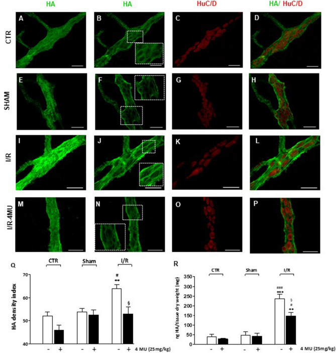
In vivo 4-MU treatment regulates HA levels in rat small intestine LMMPs after I/R injury. (A–P) Representative confocal microphotographs showing HA staining of the rat small intestine myenteric plexus in control conditions (CTR, A–D), in sham-operated animals (E–H) after ischemia followed by 24 h of reperfusion (I/R, I–L) and in the I/R group treated with 4-methylumbelliferone (4-MU, 25 mg/kg, ip) (M–P). In all groups, HA intensely stained the surface of the myenteric ganglia and interconnecting fibers (panels A, E, I, M). In myenteric ganglia median sections, HA immunofluorescence was prevalently found in neuronal soma and in the perineuronal space (insets, panels B, F, J, N), as demonstrated by double-staining with the pan-neuronal marker, HuC/D. Bar 50 μm. (Q) HA density index of HA in LMMP preparations obtained the different experimental groups with and without treatment of 4-MU, as indicated on bottom of bars. Data are reported as mean ± SEM. **P < 0.001 vs. CTR, #P < 0.05 vs. sham-operated, §P < 0.05 vs. I/R by one-way ANOVA with Tukey’s post hoc test, N = 5 rat/group. (R) Quantification of HA levels by ELISA assay in small intestine LMMP preparations obtained from the different experimental groups with and without treatment of 4-MU, as indicated on bottom of bars. HA levels are expressed as ng of HA normalized per mg of dry tissue weight. ***P < 0.001 and **P < 0.01 vs. CTR; ###P < 0.001 and #P < 0.05 vs. sham-operated by one-way ANOVA with Tukey’s post hoc test, N = 6 rat/group.

