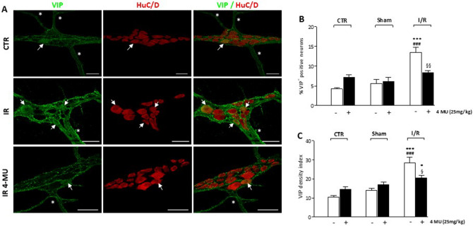
Figure 8.
4-MU modulation of VIPergic innervation in the rat small intestine after I/R injury. (A) Representative confocal microphotographs showing the distribution of VIP (green) and HuC/D (red) in rat myenteric plexus obtained from CTR, I/R and I/R-4MU groups (N = 5 rats per group). Scale bars = 50 μm. Arrows indicate VIP+-HuC/D+ neurons, asterisk interconnecting fibers. (B) Percentage of VIP+-HuC/D+ neurons with respect to total HuC/D+ neurons and (C) density index of VIP immunoreactivity in small intestine LMMP whole-mount preparations in the different experimental groups (N = 5 rats per group). Data are reported as mean ± SEM. Difference significance: **P < 0.01, ***P < 0.001 vs. CTR; ##P < 0.01 vs. sham-operated; §§P < 0.01 vs. I/R by one way ANOVA.

