
Figure 2.
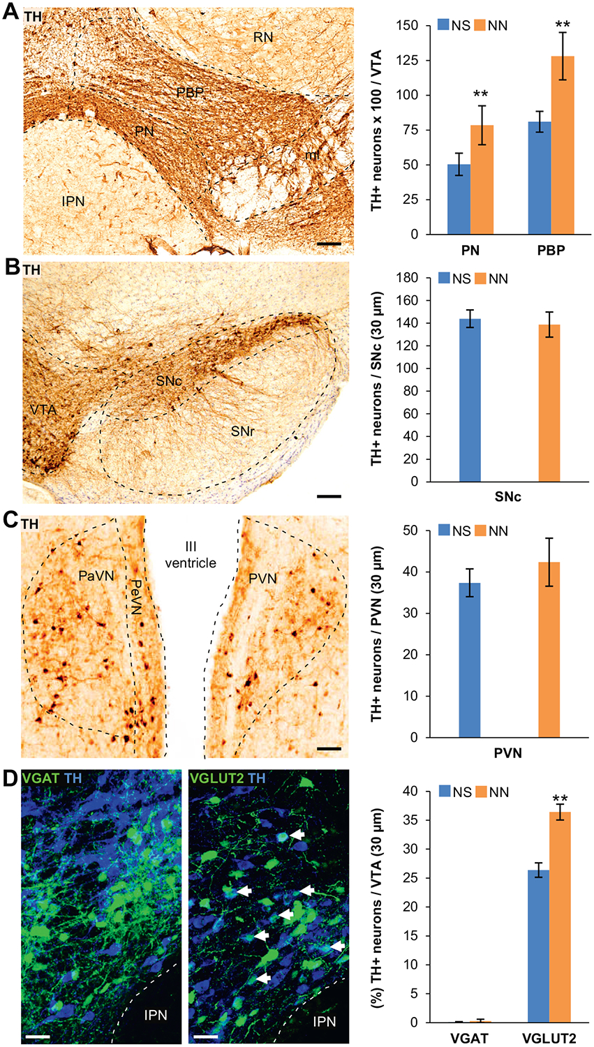
Neonatal nicotine (NN)-treated mice display an increased number of tyrosine hydroxylase (TH)1 neurons in the ventral tegmental area (VTA) after adult nicotine (AN) consumption. (A) Representative image of DAB immunoreactivity of the VTA labeled with TH marker after AN exposure. DAB stereological quantification shows that after AN exposure, the number of TH+ neurons per VTA section (30 μm) in paranigral (PN) (n = 6 mice per group, t10 = 3.71, p, .01) and parabrachial pigmented (PBP) (n = 6 mice per group, t10 = 3.39, p, .01) subnuclei of the VTA is significantly increased in NN-treated mice compared with control mice (neonatal saline [NS]). Graph shows mean 6 SE. **p, .01. Scale bar = 100 mm. (B, C) Representative images of TH-DAB immunostaining performed on the substantia nigra compacta (SNc) (B) and paraventricular nuclei (PaVN) (C) after AN exposure. Stereological quantification of DAB TH-immunoreactivity (graphs) shows that NN treatment does not affect the number of TH1 neurons in other dopaminergic regions of the brain. Graph shows mean 6 SE. Scale bars = 100 mm. (D) Confocal images showing TH and green fluorescent protein (GFP) immunofluorescence in the VTA of NN/AN-treated VGAT-Cre (left) and VGLUT2-Cre (right) mice injected with Cre-dependent green fluorescent protein–adeno-associated virus reporter. Epifluorescence quantification (%) of TH1/GFP1 neurons following AN exposure shows a significant increase in TH expression within glutamatergic neurons (arrows) in NN-treated mice (n = 6 mice per group, t10 = 25.43, p, .01). Graph shows means 6 SE. **p, .05. Scale bars = 50 mm. IPN, interpeduncular nucleus; PVN, periventricular nucleus; RN, red nucleus; SNr, substantia nigra reticulata.