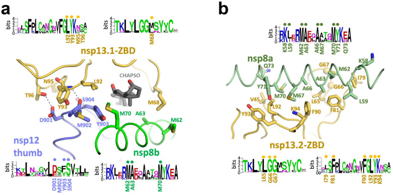
Figure 3. Interactions of the nsp13-ZBDs with the RTC.
a.-b. Views of the nsp13-ABD:RTC interactions. Proteins are shown as α-carbon backbone worms. Side chains that make protein:protein interactions are shown. Polar interactions are shown as dashed grey lines. Sequence logos (Schneider and Stephens, 1990) from alignments of α- and β-CoV clades for the interacting regions are shown. The residues involved in protein:protein interactions are labeled underneath the logos. Dots above the logos denote conserved interacting residues.
a. View of the tripartite nsp13.1-ZBD:nsp8b-extension:nsp12-thumb interaction. The adventitiously-bound CHAPSO molecule is shown in dark grey.
b. View of the nsp13.2-ZBD:nsp8a-extension interaction.
See also Figure S6.

