Fig. 2.
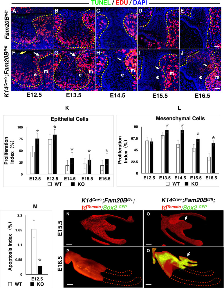
Cell fate change and ectopic renewal of Sox2(+) cells in the dental epithelium of K14Cre/+;Fam20Bfl/fl mice. A–M GAG deficiency in the dental epithelium led to ectopic over-proliferation and less apoptosis in the native incisors. EdU incorporation analysis showed ectopic over-proliferation (white arrows in F–J) in the GAG-deficient dental epithelium (e, plotted by dashed lines) and dental mesenchyme (m) at the mesial/lingual side of native incisors in the presumptive location of supernumerary teeth formation starting at E13.5 compared to the normal control mice (2-sample t tests, P < 0.05). TUNEL assay revealed reduced apoptosis (yellow arrow) in GAG-deficient dental epithelium at E12.5 (2-sample t tests, P < 0.01). N–Q Three-dimensional reconstruction of lower incisor confocal images after tissue clearing of mandibles. tdTomato indicates the dental epithelium. GFP indicates the Sox2(+) cells. On the sagittal sections of E15.5 incisors (N, O), an ectopic thickening of dental epithelium was identified at the lingual side of the native enamel organ in K14Cre/+;Fam20Bfl/fl mice (arrow) compared to the normal control. The slight bulge (white arrow) at the lingual side of the enamel organ in the Fam20B-mutant mice indicates the initiation of the supernumerary tooth outgrowth. At E16.5 (P, Q), Sox2(+) cells showed ectopic renewal in the Fam20B-deficient native enamel organ and actively contributed to the development of successive enamel organ (arrow). In contrast, the normal controls had lost Sox2 expression at this stage. The dashed lines in (P) and (Q) outlined the remaining parts of the enamel organs that were not able to be displayed due to the working distance of the confocal. Scale bars: 25 μm in (A–J); 50 μm in (N–Q)

