Fig. 1.
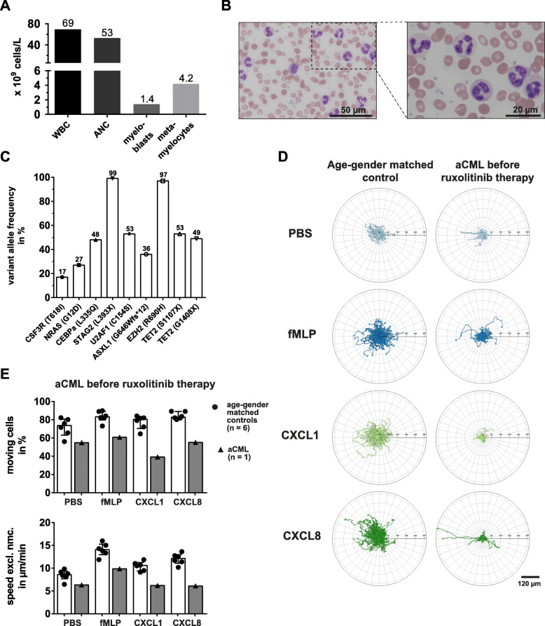
Migration of aCML neutrophils is severely impaired. a Peripheral blood parameters of the untreated aCML patient. b Microscopic images of peripheral blood smear from a 69-year old, untreated aCML patient. 50x magnification (top) and 100x magnification (bottom) are shown. c Variant allele frequency (VAF) of mutated candidate genes in unseparated patient peripheral blood leukocytes. d Representative trajectory plots of migrating neutrophils from an age- and gender-matched control (left) and the aCML patient before therapy (right). From top to bottom, the cells were treated with PBS as a vehicle control, fMLP [10 nM], CXCL1 [100 ng/ml], and CXCL8 [100 ng/ml] in vitro, continuously imaged for 1 h under a widefield microscope and single cells were automatically tracked. e Statistical summary of percentage of moving cells (top) and speed excluding non-moving cells (speed excl. Nmc., bottom) of the untreated aCML patient (black triangles, grey bars) and the age- and gender-matched controls (black dots, white bars; n = 6). Each symbol represents a single individual. Bars are given as median ± interquartile range

