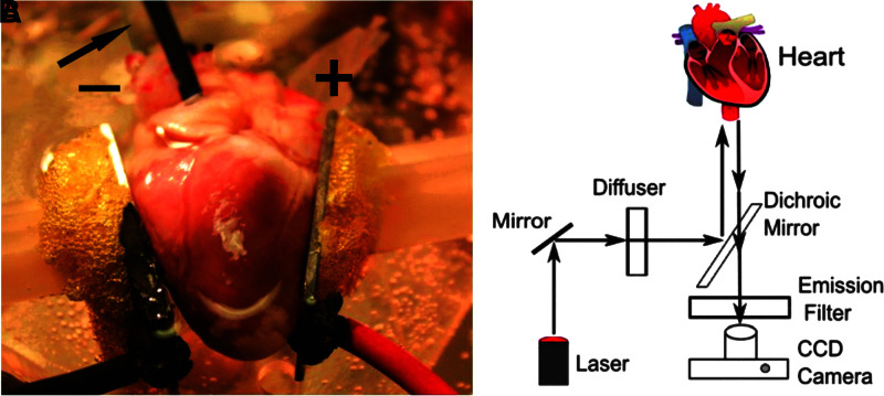
FIG. 1.
Experimental setup for ex vivo defibrillation studies and optical mapping setup. (A) Heart preparation. An excised rabbit heart is perfused and superfused with Tyrode's solution. Aluminum plate electrodes, labeled “+” (anode) and “−” (cathode), are placed in contact with the heart on the left and right ventricles. The arrow is pointing to the transparent cannula that perfuses the heart; the black tube next to the cannula is inserted through the left atrial wall into the left ventricle to provide an outlet from the left ventricle (since the aorta is cannulated). The yellow foamy substance between the electrodes and the (white) manipulators is water-insoluble glue used for mounting the electrodes. (B) Optical mapping setup. Laser light is diffused and directed toward the heart to excite the fluorescent dye. The fluorescent light then passes the dichroic mirror and an emission filter and is recorded by a CCD camera. CCD, charge-coupled device.

