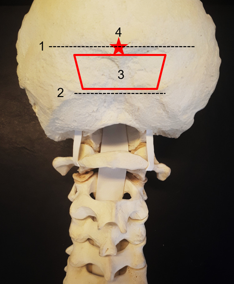
Figure 1.

(1) Superior nuchal line—insertion of the trapezius, splenius capitis, and sternocleidomastoid muscle. The transverse sinus and the torcula are internal to this line. (2) Inferior nuchal line—insertion of the obliquus capitis superior, rectus capitis posterior major, and rectus capitis posterior minor muscle. We generally perform, when necessary, decompression below this area. (3) The “red trapezium” corresponds to the area of preference to insert the occipital screws—just below the superior nuchal line to avoid a prominent plate, in the midline, where the occipital squama is thicker. (4) The “red star” is the external occipital protuberance, which corresponds to the confluence of the dural sinuses (torcula).
