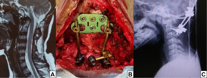
Figure 4.
This patient underwent a posterior fossa decompression and C1-2 wiring 3 years before being referred to our center. He developed severe pain when flexing the head, along with dysphagia. In (A), a sagittal T2 MRI showing the dens compressing the spinal cord and posterior fossa decompression. In (B), an occipital plate was affixed with 4 screws just below the superior nuchal line. On the axis, there were 3 screws: bilateral pars screws and a unilateral laminar screw. (C) Postoperative lateral plain radiograph (C).

