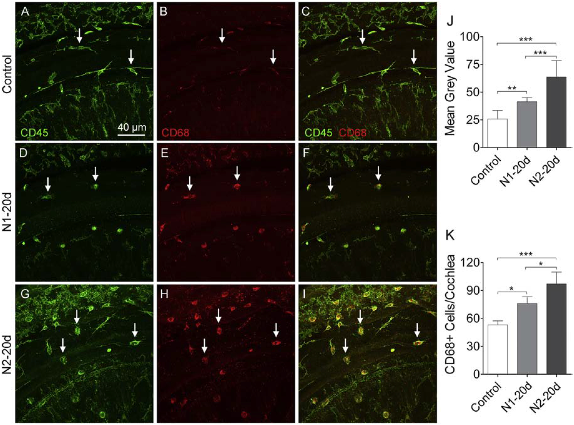
Figure 13. CD68 immunoreactivity in basilar membrane macrophages in control and noise-damaged cochleae.

A-C. Macrophages in a control cochlea display weak CD68 immunoreactivity. D-F. 20 days after the initial noise exposure, the number of CD68-positive cells increases along with their immunoreactivity. G-I. 20 days after the second noise exposure, there is a further increase in the number of CD68-positive cells as well as their immunoreactivity. J. Comparison of the immunoreactivity of CD68-positive cells in the control, N1–20d, and N2–20d group. CD68 immunoreactivity is elevated at 20 days after the first noise exposure and further increases after the second noise exposure. K. Comparison of the number of CD68-positive cells on the basilar membrane per cochlea in the control, N1–20d, and N2–20d group. The number of CD68-positive cells increases at 20 days after the initial noise exposure and further increases after the second noise exposure. n = 4 cochleae biological replicates for each group. (*, ** and *** indicate P < 0.05, 0.01 and 0.001, respectively).
