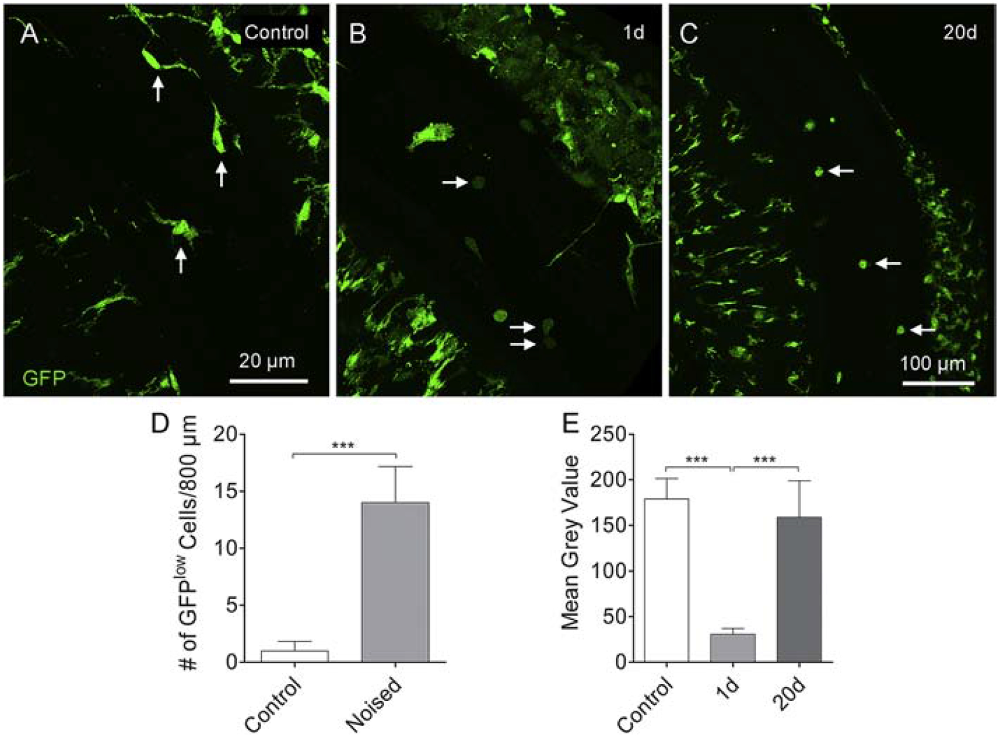
Figure 8. Shift from GFPl°w to GFPhigh monocyte-like cells in Cx3cr1GFP/+ cochleae after noise exposure.

A. Tissue macrophages on the basilar membrane are GFPhigh under resting conditions, and the intensity of the fluorescence is relatively homogenous across GFP-positive cells (arrows). B. One day after noise exposure, GFPl°w cells emerge on the surface of the basilar membrane as well as in the lateral wall and osseous spiral lamina. These cells have a monocyte shape and size (arrows). C. Small, round monocyte-like GFPhigh cells are present in the cochlea examined at 20-days after noise exposure (arrows). D. Comparison of the number of GFPl°w cells between control cochleae and cochleae examined at 1-day after noise exposure. The number of GFPl°w cells in the noise-damaged cochleae is significantly higher than that in the control cochleae. n = 4 cochleae biological replicates for each group. E. Comparison in the GFP fluorescence intensity of basilar membrane macrophages between the control, 1-day and 20-day groups (*** indicates P < 0.001).
