
Fig. 2.
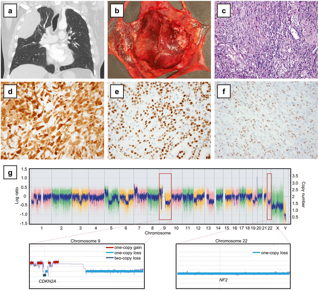
BAP1-mutant localized pleural mesothelioma (Case 1). a Computed tomography demonstrated a circumscribed pleural mass in the right upper lobe. b Grossly, the tumor was solitary with no evidence of serosal spread. c The tumor showed biphasic histology with both epithelioid and sarcomatoid components, histologically indistinguishable from diffuse malignant pleural mesothelioma (hematoxylin and eosin, 200×). By immunohistochemistry, tumor cells expressed calretinin (d) and WT1 (e) and showed complete loss of nuclear BAP1 expression (f), with positive control BAP1 staining in the background stromal and inflammatory cells. g Single nucleotide polymorphism array analysis (top) showed homozygous deletion of CDKN2A at 9p (close-up: bottom left) and hemizygous deletion of NF2 at 22q (close-up: bottom right)