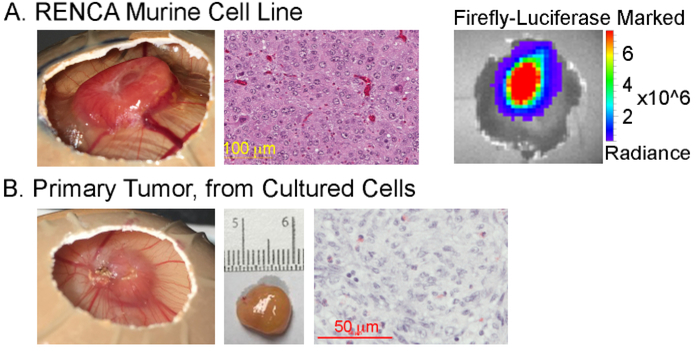
Figure 2: Representative tumor development from clear cell renal cell carcinoma.
Tumors resulting from implantation of (A) the murine renal cell carcinoma cell line, RENCA, or (B) cultured cells derived from a digested human primary tumor. The measurement scale adjacent to the excised tumor in (B) shows 1 mm markings. Corresponding histology in (A) and (B) shows hematoxylin and eosin staining. In (A), the representative bioluminescence imaging of firefly-luciferase-marked RENCA cells is also shown.

